Transphenoidal Removal for Pituitary Tumour
The pituitary gland is a pea-sized structure located at the base of the brain. It functions by producing hormones that control or regulate various functions of the body such as growth, metabolism, sexual development and reproduction.
The pituitary gland releases hormones that act directly on the body tissues and also regulates the production of hormones from other glands such as the thyroid and adrenal glands. Thus, pituitary tumours can lead to overproduction of one or more hormones causing conditions such as hyperthyroidism, gigantism, Cushing’s syndrome and abnormal discharge from the nipples of the breasts. As they grow they may put pressure on the optic nerve affecting vision. Their diagnosis is thus made on the basis of endocrine testing for hormone levels. The diagnosis is further confirmed by imaging of the head (MRI) and visual field testing.
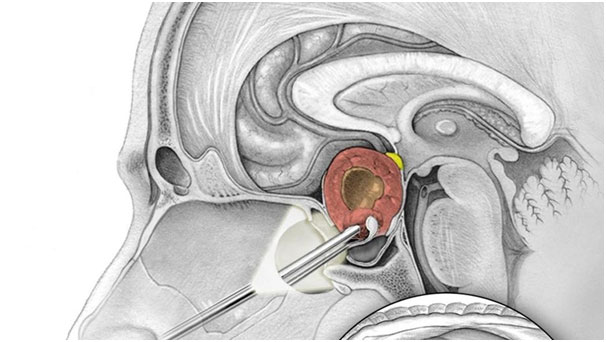
Transnasal transsphenoidal surgery is a minimally invasive technique performed to remove pituitary adenomas by inserting an endoscope through the nose. An endoscope is a long tube with a camera attached at the end that sends images to a computer screen for the surgeon to view inside the body.
INDICATIONS
Endoscopic transnasal transsphenoidal surgery can be utilised to remove pituitary tumours which are compressing critical brain structures or are over secreting certain hormones.
SURGICAL PROCEDURE
The surgery is performed under general anaesthesia by your neurosurgeon and ear, nose and throat (ENT) surgeon.You will lie on your back on the operating table and your nasal cavity will be prepared with antibiotic and antiseptic solution.An image-guided device is placed on your head which creates a 3D map on a computer screen. This map assists your surgeon in navigating through the nose.
A thin endoscopic tube attached to a lighted device and a video camera at its end is inserted through one nostril and moved up to the back of the nasal cavity. A small portion of the nasal septum separating the two nostrils and the wall of the sphenoid sinus is opened.
The surgeon then makes an opening in the thin bone overlying the pituitary gland called the sella, to view the dura (the covering of the brain). The dura is then opened to view the tumour and pituitary gland.Your surgeon uses special instruments called curettes through the other nostril to remove the tumour.
At the conclusion of the procedure your surgeon may take a fat graft from your abdomen or leg which is used to repair the opening into the skull base and to prevent any CSF fluid leakage. If there has been a spinal fluid leak, your surgeon may apply biologic glue over the fat graft to prevent leakage of cerebrospinal fluid into the nasal cavity.
POST-OPERATIVE COURSE
After the surgery, medications are prescribed to control nausea, pain and nasal congestion. Your doctor will check to see if the pituitary gland is functioning normally. Hormonal medications may be prescribed if the pituitary gland fails to produce the required level of hormones.
Avoid coughing, sneezing, blowing your nose, and straining during bowel movements for a few weeks after surgery. Crusts can form in your nose, causing nasal congestion. Nasal saline rinses may be recommended to remove the crusts and promote healing of the wounds. Resume normal activities gradually.
If a fat graft has been obtained from the abdomen or leg, there will be a wound with dissolvable stitches. It is important to keep it clean and dry.
RISKS AND COMPLICATIONS
As with any surgery, pituitary surgery can be associated with certain risks and complications, which may include:
- Loss of vision due to optic nerve injury
- Pituitary gland dysfunction
- Nasal deformity and bleeding
- Infection
- CSF leak
- Salt and water imbalance